The Normal Adrenal Glands on Ultrasound Are Best Described as
Presentation1 Pptx Ultrasound Examination Of The Adrenal Glands And

14 Adrenal Glands Radiology Key

14 Adrenal Glands Radiology Key

Pin On Echo
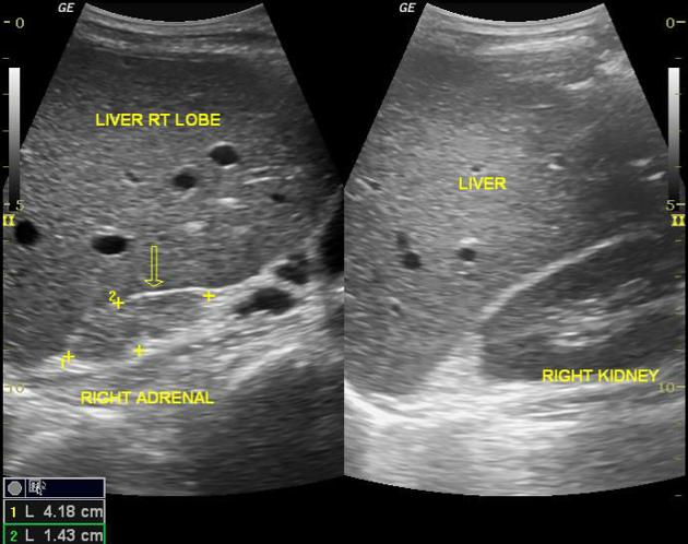
Abdomen And Retroperitoneum 1 10 Adrenal Glands Case 1 10 1 Normal Adrenals Ultrasound Cases
Abdomen And Retroperitoneum 1 10 Adrenal Glands Case 1 10 1 Normal Adrenals Ultrasound Cases

14 Adrenal Glands Radiology Key

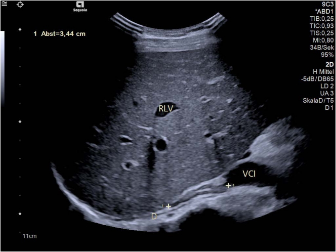
Abdomen And Retroperitoneum 1 10 Adrenal Glands Case 1 10 1 Normal Adrenals Ultrasound Cases

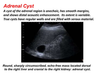
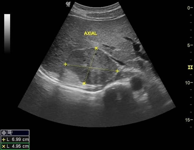
Adrenal Endothelial Cyst With A Preoperative Image Suggesting Malignancy Endocrinologia Y Nutricion English Edition

Ultrasound Examination Hypoechoic Mass 43 40 Mm In The Left Adrenal Gland Download Scientific Diagram
Pancake Adrenal Gland In Patient With A Right Pelvic Kidney Radiology Case Radiopaedia Org
Ultrasonography

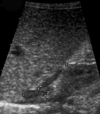
Ultrasonography Of The Right Adrenal Gland Showing An Ovoid Tumour Download Scientific Diagram

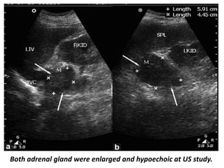
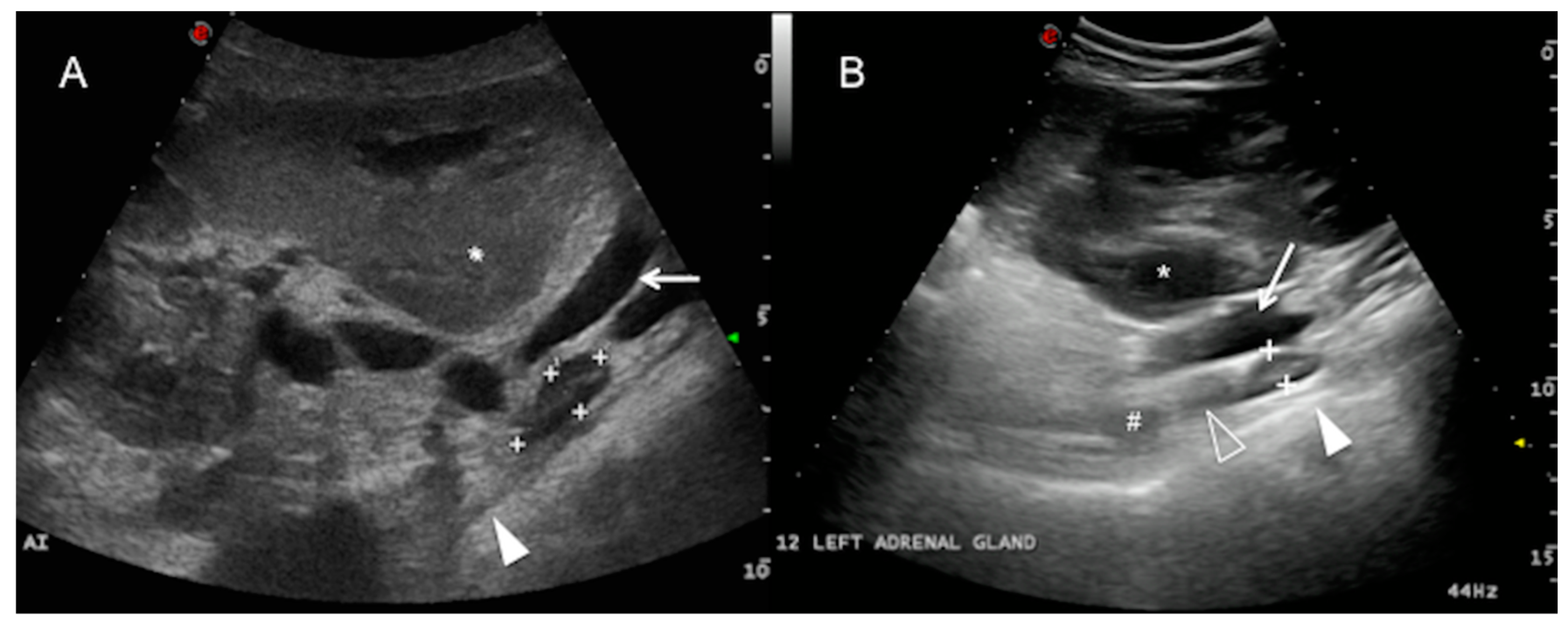
A Case Of Diffuse Enlargement Of The Right Adrenal Gland Due To Download Scientific Diagram
Presentation1 Pptx Ultrasound Examination Of The Adrenal Glands And
Animals Free Full Text Ultrasonography Of Normal Adrenal Glands In Adult Holstein Friesian Cows A Pilot Study Html
The Adrenal Glands Radiology Key

14 Adrenal Glands Radiology Key
Adrenal Pheochromocytoma Radiology Case Radiopaedia Org
Liver And Adrenal Gland Trauma Radiology Case Radiopaedia Org


Comments
Post a Comment